PRODUCT CENTER
特色產(chǎn)品當(dāng)前位置:首頁(yè) > 特色產(chǎn)品
真核細(xì)胞驅(qū)動(dòng)蛋白馬達(dá)蛋白利用 ATP 水解的能量沿細(xì)胞骨架微管網(wǎng)絡(luò)移動(dòng)貨物,如染色體和囊泡 。它們?cè)诩?xì)胞內(nèi)運(yùn)輸?shù)姆椒矫婷娑及l(fā)揮著重要作用,并廣泛參與各種生理過(guò)程,包括胚胎發(fā)育、軸突運(yùn)輸和細(xì)胞分裂。許多驅(qū)動(dòng)蛋白在細(xì)胞分裂中起著重要作用,以及驅(qū)動(dòng)蛋白的過(guò)度表達(dá)與癌癥(如視網(wǎng)膜母細(xì)胞瘤)有關(guān)的事實(shí),使它們成為開(kāi)發(fā)抗有絲分裂藥物非常有潛力的靶標(biāo)。許多驅(qū)動(dòng)蛋白僅在細(xì)胞分裂期間表達(dá)的證據(jù)也表明,它們可能優(yōu)于當(dāng)前的抗有絲分裂藥物靶標(biāo),例如普遍表達(dá)的Tubulin。
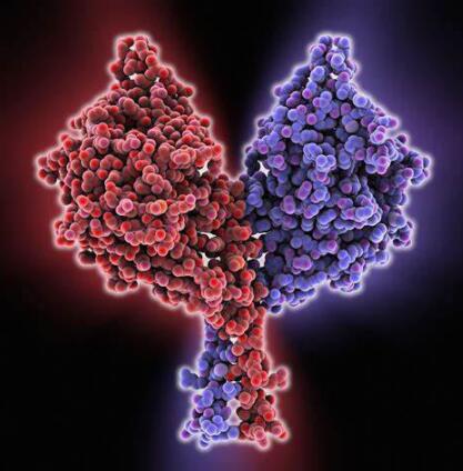
驅(qū)動(dòng)蛋白的一般結(jié)構(gòu):
驅(qū)動(dòng)蛋白分類和結(jié)構(gòu)多樣性
迄今為止,已經(jīng)在從酵母(釀酒酵母 含有 6 種驅(qū)動(dòng)蛋白)和真菌到人類(目前有13種驅(qū)動(dòng)蛋白)等物種中鑒定了大約 150 種驅(qū)動(dòng)蛋白。分類基于馬達(dá)蛋白域的同源性。目前一致認(rèn)為至少有九類驅(qū)動(dòng)蛋白,驅(qū)動(dòng)蛋白分類如表1所示,驅(qū)動(dòng)蛋白命名(在表 1 中以粗體顯示)基于驅(qū)動(dòng)蛋白內(nèi)馬達(dá)蛋白結(jié)構(gòu)域的位置,因此 N-蛋白質(zhì)在蛋白質(zhì)的氨基末端包含一個(gè)運(yùn)動(dòng)結(jié)構(gòu)域,而 C- M-馬達(dá)蛋白分別位于羧基末端和蛋白質(zhì)中間。
考慮到不同類驅(qū)動(dòng)蛋白的馬達(dá)之間相對(duì)較高的百分比差異,很可能識(shí)別類特異性的驅(qū)動(dòng)蛋白藥物,這一結(jié)構(gòu)證據(jù)得到了生化數(shù)據(jù)的支持,這些數(shù)據(jù)表明不同類別的果蠅驅(qū)動(dòng)蛋白表現(xiàn)出對(duì)各種 ATP 類似物的不同的利用效率。實(shí)驗(yàn)表明,即使在同一的物種內(nèi),不同驅(qū)動(dòng)蛋白之間也存在明顯的結(jié)構(gòu)差異,可用于選擇性藥物開(kāi)發(fā)。
表 1:驅(qū)動(dòng)蛋白超家族蛋白的分類及功能
| 驅(qū)動(dòng)蛋白種類 | 細(xì)胞功能 | 候選HTS靶標(biāo)代表 | 功能性有絲分裂抑制數(shù)據(jù) | 參考文獻(xiàn) |
| N-II (BIMC) | 中心體分離 主軸動(dòng)力學(xué) (12名成員, 來(lái)自于5個(gè)門類) | AnBimC HsEg5 | 曲霉中bmc突變體抑制紡錘體分離,并引起致死表型。 在HeLa細(xì)胞中,通過(guò)顯微注射HsEg5抗體導(dǎo)致>80%的細(xì)胞有絲分裂中止。這些結(jié)果通過(guò)過(guò)表達(dá)HsEg5馬達(dá)蛋白突變體得到證實(shí)。 | 7、17、34 |
| NV (染色質(zhì)) | 通過(guò)與染色體結(jié)合并幫助它們?cè)谥衅诎迳蠈?duì)齊而參與細(xì)胞分裂。(7 名成員, 來(lái)自于3個(gè)門類) | 染色質(zhì)驅(qū)動(dòng)蛋白 | 體內(nèi)反義和體外抗體抑制及免疫耗竭實(shí)驗(yàn)證明了染色質(zhì)驅(qū)動(dòng)蛋白在紡錘體組織和染色體定位中的重要作用。運(yùn)動(dòng)抑制導(dǎo)致爪蟾細(xì)胞有絲分裂阻滯和細(xì)胞死亡。人類染色體驅(qū)動(dòng)蛋白的異常與視網(wǎng)膜母細(xì)胞瘤有關(guān) | 18、19、20、21 |
| N-VII | 參與中期板上的染色體聚集,并可能參與后期 A 的染色體運(yùn)動(dòng) (2 個(gè)成員,來(lái)自于1個(gè)門類) | HsCENP-E | 無(wú)馬達(dá) CENP-E 蛋白在人源細(xì)胞系體內(nèi)過(guò)表達(dá)導(dǎo)致染色體無(wú)法在中期板上對(duì)齊并導(dǎo)致有絲分裂阻滯。此外,將 CENP-E 抗體顯微注射到 HeLa 細(xì)胞中也會(huì)導(dǎo)致有絲分裂阻滯,持續(xù) 4 到 17 小時(shí),并導(dǎo)致所有細(xì)胞進(jìn)入細(xì)胞凋亡。 | 22、23、24、25 |
| N-VI (MKLP1) | 參與有絲分裂的后期 B 和胞質(zhì)分裂。 (5 個(gè)成員,來(lái)自于2個(gè)門類) | HsMKLP1 | N-VI 家族的果蠅突變體表明,這種馬達(dá)蛋白對(duì)于胞質(zhì)分裂至關(guān)重要。 | 26、27 |
| C (C端) | 可能通過(guò)調(diào)節(jié)微管動(dòng)力學(xué)參與有絲分裂和減數(shù)分裂紡錘體的形成。一些成員可能是專門的囊泡轉(zhuǎn)運(yùn)蛋白。 (18名成員,來(lái)自于7個(gè)門類) | HsKifC3 | 低等真核生物(酵母)中的突變分析表明,此類蛋白質(zhì)中的無(wú)效突變體導(dǎo)致 G2/M 的致命有絲分裂阻滯。 | 35、36、37、38、39 |
| M (MCAK/KIF2 ) | 參與后期染色體運(yùn)動(dòng)和微管動(dòng)力學(xué)。一些成員是囊泡驅(qū)動(dòng)蛋白。(10名成員,來(lái)自于4個(gè)門類) | HsMCAK | 反義抑制哺乳動(dòng)物的M蛋白導(dǎo)致后期染色體分離的中斷。一個(gè)無(wú)馬達(dá)蛋白突變體的過(guò)表達(dá)也導(dǎo)致了后期的破壞。 | 6、32、33 |
| N-I (KHC) | 細(xì)胞器/囊泡運(yùn)輸 (15名成員,來(lái)自于7個(gè)門類) | HsKHC | 在有絲分裂中沒(méi)有作用 | 30,31 |
| N-III (Unc104) | 細(xì)胞器/囊泡運(yùn)輸,特別是突觸囊泡和線粒體 (18名成員,來(lái)自于4個(gè)門類) | HsKIF1C | 在有絲分裂中沒(méi)有作用 | 28 |
| N-Ⅳ (KRP85/95) | 細(xì)胞器/囊泡運(yùn)輸特別是順行囊泡運(yùn)輸。 (13 名成員, 來(lái)自于4個(gè)門類) | HsKIF3C | 在有絲分裂中沒(méi)有作用 | 29 |
用于 HTS 分析的驅(qū)動(dòng)蛋白靶標(biāo)的選擇
從上表我們可以看到,驅(qū)動(dòng)蛋白的細(xì)胞功能主要可分為兩大類:膜囊泡和細(xì)胞器的運(yùn)輸和定位,以及有絲分裂紡錘體的形態(tài)發(fā)生和染色體運(yùn)動(dòng) [12],九類驅(qū)動(dòng)蛋白中有四類專門參與細(xì)胞分裂(表 1 的黃色/綠色陰影區(qū)域),兩類(C- 和 M-)同時(shí)包含囊泡轉(zhuǎn)運(yùn)蛋白和有絲分裂馬達(dá)蛋白(表 1 的淺黃色陰影區(qū)域),三個(gè)類別(NI、NIII 和 N-IV)僅在囊泡運(yùn)輸中起作用(表 1 的無(wú)陰影區(qū)域)。
我們從每個(gè)驅(qū)動(dòng)蛋白類中選擇了一個(gè)人類同源蛋白[6,17,21,22,26,28,29,30,39],從Aspergillus(一種人類病原體)中選擇了兩個(gè)真菌驅(qū)動(dòng)蛋白[34], 一個(gè)人類病原體作為 HTS 檢測(cè)的基礎(chǔ)(這些顯示在表 1 中)。通過(guò)用化合物庫(kù)篩選每種蛋白質(zhì),可以生成一個(gè)初級(jí)化合物庫(kù),該化合物庫(kù)應(yīng)該允許識(shí)別選擇性靶向細(xì)胞分裂特定類別的人類驅(qū)動(dòng)蛋白(表 1 的深色陰影區(qū)域)的化合物,同時(shí)對(duì)囊泡轉(zhuǎn)運(yùn)驅(qū)動(dòng)蛋白沒(méi)有影響。這種化合物很有可能是治療人類疾病如癌癥的有效的抗有絲分裂劑。同樣,可以將與曲霉驅(qū)動(dòng)蛋白特異性反應(yīng)的化合物用作潛在的抗真菌藥物。
驅(qū)動(dòng)蛋白為什么能作為抗有絲分裂藥物篩選靶點(diǎn)?
在開(kāi)始一項(xiàng)昂貴的藥物開(kāi)發(fā)計(jì)劃之前,必須嚴(yán)格評(píng)估候選蛋白的適用性,以滿足有效藥物靶標(biāo)的標(biāo)準(zhǔn)。驅(qū)動(dòng)蛋白將成為抗有絲分裂藥物開(kāi)發(fā)的絕佳靶點(diǎn)的證據(jù)來(lái)自一系列實(shí)驗(yàn)方法:包括突變分析 [40]、抗體抑制實(shí)驗(yàn) [7,32] 和驅(qū)動(dòng)蛋白活性的反義抑制 [19]。這些證據(jù)總結(jié)在表 1 中,可以看出特定有絲分裂特異性驅(qū)動(dòng)蛋白的功能性抑制導(dǎo)致細(xì)胞分裂的抑制。
在低等真核生物中,突變分析有助于闡明有絲分裂驅(qū)動(dòng)蛋白的作用。單個(gè)驅(qū)動(dòng)蛋白的突變通常會(huì)導(dǎo)致有絲分裂阻滯和致死表型。我們選擇作為抗真菌靶點(diǎn)的 AnBimC 蛋白就是這種情況[34]。然而,在某些情況下,兩個(gè)高度同源的基因執(zhí)行相同的功能從而導(dǎo)致功能冗余[41],在這些情況下,必須同事敲除兩個(gè)基因才能產(chǎn)生有絲分裂表型變化。
重要的是,所有數(shù)據(jù)表明,針對(duì)有絲分裂驅(qū)動(dòng)蛋白的抗體會(huì)特異性影響有絲分裂過(guò)程,但對(duì)驅(qū)動(dòng)蛋白囊泡轉(zhuǎn)運(yùn)功能沒(méi)有影響 [42]。事實(shí)上,對(duì)特定驅(qū)動(dòng)蛋白的抑制通常會(huì)誘導(dǎo)一種非常具有細(xì)胞周期特異性的表型。例如,抑制 M 類驅(qū)動(dòng)蛋白 MCAK 中的運(yùn)動(dòng)區(qū)域?qū)忓N體組裝沒(méi)有影響,但確實(shí)抑制了染色體運(yùn)動(dòng) [32]。該數(shù)據(jù)表明驅(qū)動(dòng)蛋白馬達(dá)的特定抑制劑將只會(huì)特異性地抑制有絲分裂過(guò)程而不影響其他關(guān)鍵細(xì)胞功能。
驅(qū)動(dòng)蛋白靶標(biāo)相對(duì)于當(dāng)前抗有絲分裂靶標(biāo)的優(yōu)勢(shì):
微管的紡錘體蛋白Tubulin是目前抗有絲分裂藥物如紫杉醇和長(zhǎng)春堿的主要靶點(diǎn) [43,44]。人們普遍認(rèn)為,這些藥物通過(guò)在有絲分裂期間直接抑制微管動(dòng)力學(xué)發(fā)揮作用 [45,46]。由于有絲分裂細(xì)胞中的微管動(dòng)力學(xué)比靜止細(xì)胞大得多,因此對(duì)分裂細(xì)胞的特異性是有利的。藥物處理導(dǎo)致有絲分裂阻滯,在此期間細(xì)胞進(jìn)入凋亡途徑并死亡 [47]。但由于其作用機(jī)制的性質(zhì),所有抗有絲分裂劑,包括紫杉醇和長(zhǎng)春堿,都會(huì)在某種程度上對(duì)正常有絲分裂細(xì)胞產(chǎn)生不利影響,例如存在于胸腺、睪丸、小腸、結(jié)腸和胎盤中的細(xì)胞 [48] .
然而,有絲分裂驅(qū)動(dòng)蛋白作為潛在的抗有絲分裂藥物靶點(diǎn)的一個(gè)主要優(yōu)點(diǎn),是它們的表達(dá)通常受到嚴(yán)格調(diào)節(jié),從而與有絲分裂事件一致。例如,HsMCAK 僅在增殖細(xì)胞中表達(dá),其表達(dá)已被證明在轉(zhuǎn)錄水平受到嚴(yán)格調(diào)控 [6];HsEg5 僅在有絲分裂期間與微管相關(guān),在體內(nèi)間期與 MT 不相關(guān) [7];染色質(zhì)驅(qū)動(dòng)蛋白僅在增殖細(xì)胞中表達(dá),在那里它們是壽命較短的蛋白質(zhì),可能受細(xì)胞周期蛋白降解機(jī)制的調(diào)節(jié) [51];研究表明,CENP-E在核破裂后立即與著絲點(diǎn)結(jié)合,并一直保持完全結(jié)合,直到B后期,當(dāng)它重新定位到B后期紡錘體,隨后通過(guò)類似周期素的途徑降解 [52]。有絲分裂驅(qū)動(dòng)蛋白表達(dá)的嚴(yán)格調(diào)控預(yù)示著這些將是高度特異性的抗有絲分裂靶點(diǎn),具有最小的劑量限制副作用。
產(chǎn)品推薦:
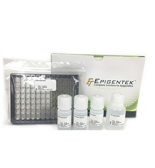
在這里給大家?guī)?lái)一款驅(qū)動(dòng)蛋白活性檢測(cè)試劑盒,可用于篩選影響驅(qū)動(dòng)蛋白活性的藥物。
產(chǎn)品詳情參見(jiàn):驅(qū)動(dòng)蛋白(Kinesin)活性檢測(cè)試劑盒
| 產(chǎn)品名稱 | 貨號(hào) |
| Kinesin ELIPA Kit | BK060 |
參考文獻(xiàn):
Wang, SZ & Alder, R. Chromokinesin: a DNA-binding, kinesin-like nuclear protein. J. Cell Biol. 128: 761-768 [1995]
Hughes, D. Predicting the future for R&D - science or art? Drug Disc. Today 3: 487-489 [1998]
Krantz, A. Diversification of the drug discovery process. Nature Biotech. 16: 1294 [1998]
Rao, K. Short Communication. Drug Disc. Today. 3: 349 [1998]
Ansell, J. Hot and cold areas of therapeutic R&D: A survey of the top 50 pharma companies. Mod. Drug Disc. May/June: 19-22 [1999]
Endow, S., Henikoff, S., Niedziela, L. Mediation of meiotic and early mitotic chromosome segregation in Drosophila by a protein related to kinesin. Nature 345:81-83 [1990]
Endow, S., Chandra, R., Komma, D., Yamamoto, A., Salmon, E. Mutations of the Drosophila ncd microtubule motor protein cause centrosomal and spindle pole defects in mitosis. J. Cell Sci. 107:859-867 [1994]
Meluh, P., Rose, M. KAR3, a kinesin-related gene required for yeast nuclear fusion. Cell 60:1029-1041 [1990]
O’Connell, M., Meluh, P., Rose, M., Morris, R. Suppression of the bimC4 mitotic spindle defect by deletion of klpA, a gene encoding a KAR3-related kinesin-like protein in Aspergillus nidulans. J. Cell Biol. 120:153-162 [1993]
Hoang, E., Whitehead, J., Dose, A., Burnside, B. Cloning of a novel C-terminal kinesin (KIFC3) that maps to human chromosome 16q13-q21 and thus is a candidate gene for Bardet-Biedl syndrome. Genomics 52:219-222 [1998]
Kim, I., Jun, D., Sohn, U., Kim, Y. Cloning and expression of human mitotic centromere-associated kinesin gene. Biochem. Biophys. Acta. 1359:181-186 [1997]
Maney, T., Hunter, A., Wagenbach, M., Wordeman, L. Mitotic centromere associated kinesin is important for anaphase chromosome segragation. J. Cell Biol. 142:787-801 [1998]
Waters, J., Salmon, E. Cytoskeleton: a catastrophic kinesin. Curr. Biol. 6:361-363 [1996]
Enos, A., Morris, N. Mutation of a gene that encodes a kinesin-like protein blocks nuclear division in A. nidulans. Cell 60:1019-1027 [1990]
Blangy, A., Chaussepied, P., Nigg, E. Rigor type mutation in the kinesin related protein HsEg5 changes its subcellular localization and induces microtubule bundling.
Sawin, K., LeGuellec, K., Phillippe, M., Mitchinson, T. Mitotic spindle organization by a plus end directed microtubule motor. Nature 359:540-543 [1992]
Blangy, A., Lane, H., d’Herin, P., Harper, M., Kress M., Nigg, E. Phosphorylation by p34cdc2 regulates spindle association of human Eg5, a kinesin related motor essential for bipolar spindle formation in vivo. Cell 83:1159-1169 [1995]
Walczak, C., Vernos, I., Mitchison, T., Karsenti, E. A model for the proposed roles of different microtubule-based motor proteins in establishing spindle bipolarity. Curr. Biol. 8: 903-913 [1998]
Vernos, I., Raats, J., Hirano, T., Heasman, J., Karsenti, E., Wylie, C. Xklp1, a chromosomal Xenopus kinesin-like protein essential for spindle organization and chromosome positioning. Cell 81:117-127 [1995]
Tokai, N., Fujimoto, A., Toyoshima, Y., Tsukita, S., Inoue, J., Yamamota, T. Kid, a novel kinesin-like DNA binding protein, is localized to chromosomes and the mitotic spindle. EMBO J. 15:457-567 [1996]
Yan, R.T., Wang, S.Z. Increased chromokinesin immunoreactivity in retinoblastoma cells. Gene 189:263-267 [1997]
Schaar, B., Chan, G., Maddox, P., Salmon, E., Yen, T. CENP-E function at kinetochore is essential for chromosome alignment. J. Cell Biol. 139:1373-1382 [1997]
Yen, T., Li, G., Schaar, B., Cleveland, D. CENP-E is a putitive kinetochore motor that accumulates just before mitosis. Nature 359:536-539 [1992]
Wood, K., Sakowicz, R., Goldstein, L., Cleveland, D. CENP-E is a plus end directed kinetochore motor required for metaphase chromosome alignment. Cell 91:357-366 [1997]
Lombillo, V., Nislow, C., Yen, T., Gelfand, V., McIntosh, R. Antibodies to the kinesin motor domain and CENP-E inhibit microtubule depolymerization-dependent motion of chromosomes in vitro. J. Cell Biol. 128:107-115 [1995]
Nislow, C., Lombillo, V.A., Kuriyamu, R., McIntosh, R. A plus end directed motor enzyme that moves antiparallel microtubules in vitro localizes to the interzone of mitotic spindles. Nature 359:543-547 [1992]
Adams, R.R., Tavares, A.A., Salzberg, A., Bellen, H., Glover, D. Pavoretti encodes a kinesin-like protein required to organize the central spindle and contractile ring for cytokinesis. Genes & Dev. 12:1483-1494 [1998].
Wright, B., Terasaki, M., Scholey, J. Roles of kinesin and kinesin-like proteins in sea urchin embryonic cell division: evaluation using antibody microinjection. J. Cell Biol. 123:681-689 [1993]
Puck TT. and Marcus PI. 1956. Action of X-rays on mammalian cells. J. Exp. Med. 103, 653-666.
Brown JM and Wouters BG, 1999. Apoptosis, p53, and tumor cell sensitivity to anticancer agents. Cancer Research, Rev, 59, 1391-1399.
Enos, AP & Morris, NR. Mutation of a gene that encodes a kinesin-like protein blocks nuclear division in Aspergillus nidulans. Cell 60: 1019-1027 [1990].
Yen, TJ., Li, G., Schaar, BT., Szilak, I. and Cleveland, DW. CENP-E is a putative kinetochore motor that accumulates just before mitosis. Nature 359(6395): 536-539 [1992].
Wang, SZ & Alder, R. Chromokinesin: a DNA-binding, kinesin-like nuclear protein. J.Cell Biol. 128:761-768 [1995].
Blangy, A., Lane, HA., d’Herin, P., Harper, M., Kress, M. and Nigg, EA. Phosphorylation by p34cdc2 regulates spindle association of human Eg5, a kinesin-related motor essential for bipolar spindle formation in vivo. Cell 83(7): 1159 [1995].
Navonne, F., Niclas, J., Hom-Booher, N., Sparks, L., Bernstein, H., McCaffrey, G and Vale, R. Cloning and expression of a human kinesin heavy chain gene: Interaction of the C-terminal domain with cytoplasmic microtubules in transfected CV-1 cells. J. Cell Biol. [1992].
Telford, EA., Wightman, P., Leek, J., Markham, AF., Lench, NJ. and Bonthron, DT. cDNA cloning, genomic organization, and chromosomal localization of a novel human gene that encodes a kinesin-related protein highly similar to mouse Kif3C. Biochem. Biophys. Res. Comm. 242(2): 407 [1998].
37)Hoang, E., Whitehead, J., Dose, A., Burnside, B. Cloning of a novel C-terminal kinesin (KIFC3) that maps to human chormosome 16q13-q21 and thus is a canidate gene for Bardet-Biedl syndrome. Genomics 52: 219 [1998].
Kim, IG., Jun, DY., Sohn, U. and Kim, YH. Cloning and expression of human mitotic centromere-associated kinesin gene. Biochim. Biophys. Acta 1359(3): 181 [1997].
Nislow, C., Lombillo, VA., Kuriyama, R. and McIntosh, JR. A plus-end directed motor enzyme that moves antiparallel microtubules in vitrolocalizes to the interzone of mitotic spindles. Nature 359(6395): 543 [1992].
Middleton, K. and Carbon, J. (1994). Kar3-encoded kinesin is a minus-end directed motor that functions with centromere binding proteins (CBF3) on an in vitroyeast kinetochore. Natl. Acad. Sci. USA. 91:7212-7216.
Aizawa, H., Sekine, Y., Takemura, R., Zhang, Z., Nangaku, M. and Hirokawa, N. (1992). Kinesin family in murine central nervous system. Cell Biol.119:1287-1296.
Sekine, Y. et al. (1994) A novel microtubule-based motor protein (KIF4) for organelle transports, whose expression is regulated developmentally. Cell Biol.127:187-201.
Enos, A.P. and Morris, N.R. (1990). Mutation of a gene that encodes a kinesin-like protein blocks nuclear division in A. nidulans. Cell60:1019-1027.
Nislow, C., Lombillo, V.A., Kuriyama, R. and McIntosh, J.R. (1992). A plus-end directed motor enzyme that moves antiparallel microtubules in vitrolocalizes to the interzone of mitotic spindles. Nature 359:543-547.
Vale, R.D., Reese, T.S. and Sheetz, M.P. (1985) Identification of a novel force generating protein, kinesin, involved in microtubule-based motility. Cell42: 39-50.
Pesavento, P.A., Stewart, R.J. and Goldstein, L.S.B. (1994). Characterization of the KLP68D kinesin-like protein in Drosophila: possible roles in axonal transport. Cell Biol.127: 1041-1048.
Wood, K., Sakowicz, R., Goldstein, L., Cleveland, D. CENP-E is a plus end-directed kinetochore motor required for metaphase chromosome alignment. Cell 91:357-366 [1997].
Ames, B.N., McCann, J. & Yamasaki, E. (1975) Methods for detecting carcinogens and mutagens with Salmonella/mammalian-microsome mutagenicity test. Mutation Res. 31, 347-364.
Shrivastava, R., John, G. W., Rispat, G., Chevalier, A., and Massingham, R. 1991. Can the in vivo maximum tolerated dose be predicted using in vitro techniques? A working hypothesis. ATLA 19: 393-402.
Berry, M.N. & Friend, D.S. (1969) High-yield preparation of isolated rat liver parenchymal cells. Journal of Cell Biology, 43, 506-520.
Schmetz, E.G., Hazelton, G.A., Hall, J., Watkins, P.B., Klaassen, C.D., & Guzelian, P.S. (1986). Induction of digitoxigenin monodigitoxoside UDP-glucuronyltransferase activity by glucocorticoids and other inducers of cytochrome P-450p in primary monolayer cultures of adult rat hepatocytes and in human liver. J. Biol. Chem., 261, 8270-8275.
作為一家具有高端的技術(shù)實(shí)力、先進(jìn)的經(jīng)營(yíng)管理水平和完善的市場(chǎng)銷售體系的生物高科技企業(yè),總部位于武漢光谷高新技術(shù)開(kāi)發(fā)區(qū),服務(wù)面向全國(guó)。艾美捷科技是集進(jìn)口試劑、實(shí)驗(yàn)室耗材銷售、技術(shù)服務(wù)與合約開(kāi)發(fā)為一體的專業(yè)化高科技公司,為用戶提供專業(yè)的前沿資訊、完備的產(chǎn)品、整合的解決方案,及優(yōu)質(zhì)的物流服務(wù)。為了更好的服務(wù)客戶,公司組建了一支經(jīng)驗(yàn)豐富的研發(fā)團(tuán)隊(duì)-艾美捷生物技術(shù)中心,進(jìn)入研發(fā)生產(chǎn)階段,將更優(yōu)質(zhì)的產(chǎn)品推薦給國(guó)內(nèi)生物領(lǐng)域的同仁們!
艾美捷科技與國(guó)內(nèi)外優(yōu)秀的生物試劑供應(yīng)商優(yōu)保持著密切的合作關(guān)系,目前已成為眾多國(guó)際知名品牌的中國(guó)總代理或一級(jí)代理,主要包括:AmyJet、AAT Bioquest、Abbexa、Abnova、Agrisera、Anogen、Biosensis、Biovision、BioVendor、Caisson Labs、Cell Biolabs、Cytoskeleton、Demeditec、Duchefa、Epigentek、Equitech-Bio、EXBio、Fitzgerald、GeneCopoeia、Hycult Biotech、ImmunoReagents、ImmunoStep、Jackson、LifeSensors、LigaTrap、Lumiprobe、Mabtech、Matreya、Norgen Biotek、Origene、ProImmune、ProSpec、ScyTek、Solis BioDyne、SouthernBiotech、StressMarq、SySy、US Biological、TRC等,可以在短時(shí)間內(nèi)為用戶提供專業(yè)的前沿資訊、完備的產(chǎn)品及物流服務(wù)。
產(chǎn)品訂購(gòu):sales@amyjet.com
郵政編碼:430070
公司地址:武漢市洪山區(qū)光谷大道35號(hào)
光谷總部國(guó)際二期時(shí)代1棟13樓
提示:本公司所有產(chǎn)品僅供科研使用,不用于臨床診斷。
版權(quán)所有:艾美捷科技有限公司 鄂ICP備10204150號(hào)-1 鄂公網(wǎng)安備:42018502004523號(hào)
第二類醫(yī)療器械經(jīng)營(yíng)備案憑證:鄂漢藥監(jiān)械經(jīng)營(yíng)備20234324號(hào)
友情鏈接:亞科因生物每日生物評(píng)論

微信掃碼在線客服